Working with Nuclear Stains
Nuclear stains are widely used in live or fixed cells to facilitate cell counts, live cell tracking, and other applications. Commonly-used nuclear stains have been verified in Vireo, and below are recommended steps to stain nuclei in fixed or live cells.
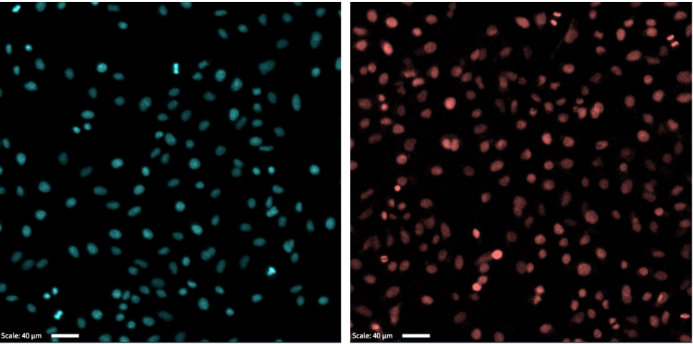
Nuclear stains with Hoechst in fixed cells (left) and SPY555-DNA (right) in live cells. White scale bar: 40um.
Fixed cells:
Step |
Function |
|---|---|
1 |
Wash cells 1x with PBS. |
2 |
Fix cells by incubating cells with 4% paraformaldehyde for 15 minutes at room temperature. |
3 |
Wash cells 3x with PBS. |
4 |
Optional: Permeabilize cells with 0.1% (v/v) Triton X-100 in PBS for 10 minutes at room temperature. |
5 |
Wash cells 3x with PBS. |
6 |
Add 1ug/ml of Hoechst 33342 to cells and incubate for 10-30 minutes. Wash 3x with PBS. |
7 |
Acquire images in the DAPI channel. Recommended imaging parameters (optimal imaging parameters to be determined empirically): Brightness (%) = 75% Exposure time (ms) = 250ms Analog Gain = 2 Digital Gain = 2 Z-cropping = Laplacian Projection = Maximum Projection |
Other nuclear stains verified in Vireo=DAPI.
Live cells:
Step |
Function |
|---|---|
1 |
Grow cells to 50% - 70% confluence. |
2 |
Dilute SPY555-DNA (Cytoskeleton CY-SC201) 1:1000 in growth media. Optional: Add verapamil to a final concentration of 10uM for better signal retention. |
3 |
Aspirate media and add SPY555-DNA/growth media from the previous step to wells. |
4 |
Incubate at 37°C in dark for at least 2 hours before still image or time-lapse acquisitions. The nuclear stain lasts 24-48 hours. |
5 |
Acquire images in the mCherry channel. Recommended imaging parameters (optimal imaging parameters to be determined empirically): Brightness (%) = 85% Exposure time (ms) = 450ms Analog Gain = 2 Digital Gain = 4 Z-cropping = Laplacian Projection = Maximum Projection |